Chirality – when the mirror self is different

All you sci-fi enthusiasts out there will be familiar with the common trope of the mirror self; a seemingly identical version of a character except that they exist in a mirrored universe. These mirror characters, in addition to being a physical mirror, are often a psychological mirror as well; evil where their original versions are good. The idea that your mirror self could have ‘opposite’ personality traits to your own, although a fun construct, runs counter to our intuition about how consequential something like a geometrical inversion should be.

Why should it matter that a person’s mole is on the other side of their face, or their hair parts in the other direction? However, there are many examples of natural objects which have mirror symmetric versions and just like in the fiction trope, this seemingly arbitrary distinction can have some surprising consequences.

What is mirror symmetry?

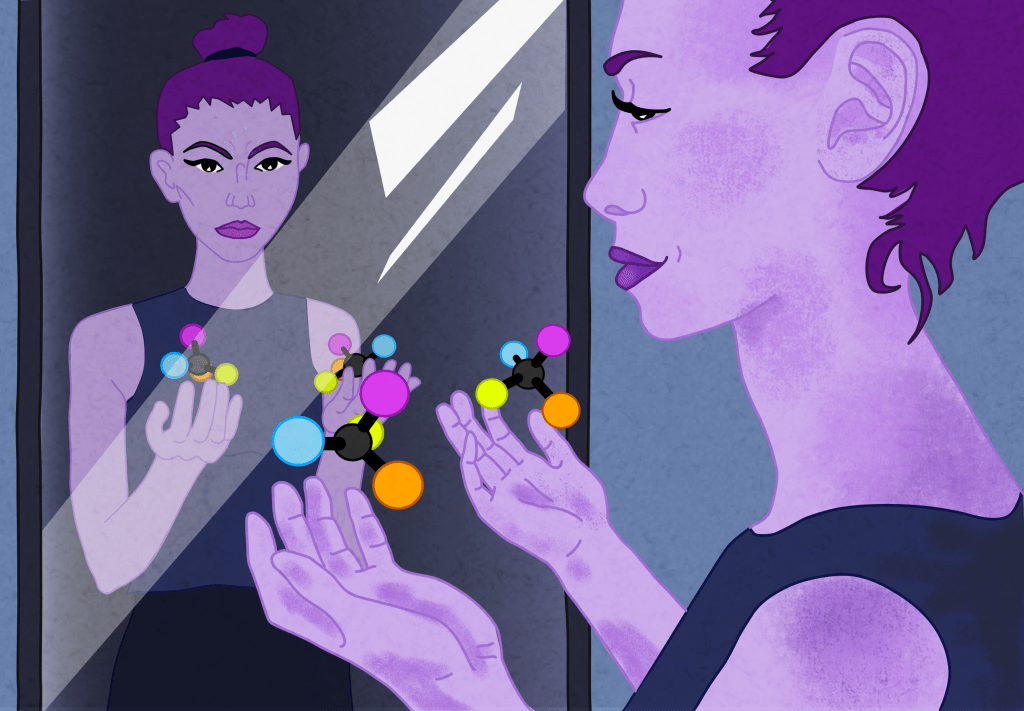
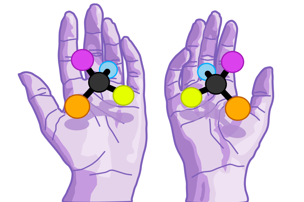
Objects which exhibit mirror symmetry are referred to as ‘chiral’. This term comes from the Greek word for hand, a clear origin, since your hands are the perfect example of two mirror symmetric objects. If you take a look at your hands, you’ll see that no matter how you turn them, you cannot lay them so they are superimposed, despite being otherwise identical. Instead, if you hold one up to a mirror, this image, which has been reflected in the plane of the mirror, will be identically orientated to the other hand.

To discriminate between the two versions we use the terms left- and right-handed. A simple way to identify which version is which is to look at the palms of your hands, if the thumb is on the left of the index finger then this is the left hand and vice versa. Scientists have similar rules which they use to identify left- and right-handed versions of the objects they’re studying.

An image of purple hands with their palms facing upwards. Above each hand is a large versions of a molecule with four different atoms each one a different colour. The molecules are mirror images of each other. They cast shadows on the hands below.
Objects like the molecules shown here come in mirror symmetric versions in the same way as the hands below.

Chirality can be seen at every level of nature, from the most fundamental aspects of physics, to macroscopic examples such as the direction that a plant’s leaves spiral around its stem. Chirality is a surprisingly important aspect to DNA, molecules that we can taste and smell, and the way that subatomic particles behave. It has also had a tragic role in the development of certain medicines. Let’s take a look at each of these examples in more detail.

Chiral spiral – the handedness of DNA

DNA is a fundamental building block of life. We all have a pretty good idea of what DNA is, and a large part of that is thanks to the determination of its shape in the mid-1900s. Now, most people would recognise the unique helical structure of DNA.

Have you ever stopped to ponder the direction of this immediately recognisable spiral though? Perhaps the handedness of the twist has never occurred to you (as pointed out by science illustration cooperative SquareCell, DNA is also often misrepresented, even in scientific media), but in fact the majority of DNA spirals in a clockwise direction, which is equivalent to being right-handed.

A gif of two arrows coloured in a gradient from green to purple which spiral away from the screen. They are mirror images reflected over an orange dotted line. Because they are mirror images, they spiral in opposite directions.
DNA can spiral in a clockwise direction, shown on the right, or anticlockwise as shown on the left. These two options are mirror images of each other. Strangely nearly every example of DNA is a clockwise spiral.

Scientists have been trying to understand the origin of this asymmetry since it was first identified. It seems that somewhere in history some mechanism created an imbalance in the relative amounts of each helical version. Most likely, this happened extremely early because the self-replicating nature of DNA means that its chirality is maintained; each new DNA molecule twists in the same direction as its parent.

Essentially, there are two potential origins of the imbalance in DNA chiralities: either the preference occurred by chance as a small difference between the two options and was magnified over time, or there exists some process that DNA participates in which always favours one handedness over the other.

If chance was the culprit, then the explanation could be as simple as basic probability; each DNA strand in the initial mixture would have had a fifty-fifty chance of being left or right-handed, just like a coin toss. If you only make a few pieces of DNA, or toss a coin a few times, you are likely to end up with a small excess of one result over the other. Alternatively, there may have been some random outside influence which created a small excess.

In either case, once a small imbalance had formed, there needed to be some reason that it was magnified, such that eventually only one handedness prevailed. There are some proposed mechanisms which could explain how this happened. However, a very interesting idea, which does not rely on a single, small excess at the beginning of life, has been under consideration for some time, waiting for a likely mechanism.

Scientists interested in the origins of chiral asymmetry in biology have long wondered if it is caused by a similar asymmetry in fundamental physics, to be discussed below. However, there hasn’t been a satisfying explanation which can link the two. In 2020, Dr Noemie Globus and Dr Roger D. Blandford at New York University published a paper suggesting a direct link between the asymmetry in the weak charge, to be discussed below, and the single spiral direction found in DNA. This is a pleasing result from an aesthetic standpoint, because it links fundamental physics to the biological world, but it has not yet been experimentally tested.

The link between the asymmetry in DNA helices and the weak charge comes from the fact that many of the mutations that allow organisms to evolve are created when a cosmic ray collides with a DNA strand. Collisions cause damage that can lead to a mutation and this mutation may be propagated to future generations if it is advantageous to the survival of the organism – the foundation of Darwin’s theory of evolution.

If cosmic rays exist in one chirality only and this chirality is more likely to damage DNA of one handedness, then it follows that this type of DNA will become dominant because the organisms it belongs to will be able to adapt more efficiently to their environments. Globus and Blandfords’ paper uses theoretical modelling to show that this theory has merit.

Philosophy of science academics will tell you though, that most of the time a good scientific theory is one which you can use to make testable predictions. The cosmic ray theory has that benefit; for example if you took two identical colonies of bacteria and subjected one to the same kind of radiation as is found in cosmic rays, you would expect to see a higher mutation rate in that colony compared to the other. I look forward to seeing the results of this experiment one day!

When mirrors smell different

Another strange example of mirror-image molecules behaving differently, occurs when they are involved in taste and smell. In this case, the peculiarity is not to do with the molecules themselves but rather how they interact with our bodies.

Taste and smell are both chemo senses meaning that what we are sensing when we smell or taste something are small quantities of molecules that are important for our health; either because they are found in our food, tell us about other humans, or they function as a warning system.

The way our bodies sense chemicals is nifty. Inside our nose, olfactory neurons in the skin lining our airways, extend their dendrites all the way down to sample the air rushing by.  On their ends are little protrusions called cilia which contain proteins that bind only to specific molecules. A binding event triggers the neuron to send a signal up through our nose, into our olfactory bulb and then to our brain where we experience the odour. Interestingly, a lot of our sense of taste also occurs in our nose because out taste buds can only discriminate between saltiness, bitterness, sweetness, sourness, and savouriness. When we eat, molecules from our food fly up the back of our throat into our nasal cavity, where they are detected in the same way as smells.

In some cases, the chemicals we sense are chiral. How we interpret the two chiralities can vary a lot; for some molecules, the two versions are exactly the same, for others they only differ in their intensity, and in others still they are interpreted as completely different. An interesting example of a molecule with two totally different tasting chiralities is the chemical carvone. The two chiral versions of carvone, referred to using the chemical notation of D and L, are present in different plants. D-carvone is found in caraway and dill, while L-carvone is found in spearmint. Pretty different tastes! Another example are the amino acids, D-amino acids usually taste sweet, whereas some L-amino acids are bitter.

Unfortunately, it is not yet fully understood what happens when chemicals interact with our chemosensory receptor proteins. Scientists suspect there is a complex combination of two processes which lead to this variable response across different chiral molecules.

The first process is the lock and key interaction which you might have learned about in school when you studied enzymes. Essentially, the enzyme or protein has a binding site that is uniquely shaped so that only one thing can fit into the space. However, if this were the sole mechanism in chemosensation then chiral molecules would either always be interpreted as different or we would only be able to sense one chirality.

Instead of being dependent on shape, the interaction could rely on the transfer of a specific amount of energy. In this case the chirality of the molecule should have no effect and both chiralities would always taste or smell the same.

Given that neither of these explanations can account for the variable way that oppositely handed molecules can smell or taste, there must be more complexity to this interaction than we first assumed. For example, a paper published in 2009 suggests that the various ways chiral molecules can be interpreted has to do with their rigidity. Counterintuitively, they found that molecules which are rigid, that is their shape can’t be ‘bent’, are those whose mirror images smell the same. On the other hand, flexible molecules seem to be those whose mirror images smell different. The interpretation for why this could work is complex, however, it’s an interesting idea which deserves further investigation.

The sad story of thalidomide

Differences in how mirror symmetric molecules smell provide a fun example of the unique ways our body can respond to chirality. However, not all examples are innocuous; when pharmaceutical drugs are involved, it can be disastrous if the two mirror image versions of that drug to behave differently in the body.

If you have had a baby or know someone who has, then you are aware that it can be an unpleasant experience, particularly at the beginning of the pregnancy. In the first trimester pregnant people often feel very nauseated and may even vomit. Often called morning sickness, this unpleasant side-effect of pregnancy is due to the change in hormone levels that occurs when conception is achieved.  

Naturally, pregnant people would prefer to avoid feeling constantly sick for three months, so anti-nausea medication has a large market. This demand was met in the late 1950s when the drug thalidomide was sold over the counter. Initially developed as an antihistamine, thalidomide turned out to be more effective at inducing drowsiness and was therefore marketed as a sleep-aid, anti-anxiety medication, and for morning sickness – you can’t feel sick if you’re asleep?

Only a few years after it was first marketed, it became obvious that thalidomide was very dangerous for developing babies. If a mother took thalidomide herself, or the baby was conceived with a father who was taking it, then the baby could be born with birth defects such as short or even absent limbs, or abnormalities to the ears, eyes, heart or gut. Even a single dose could be enough to have these impacts.

Just like other chiral molecules the two types of thalidomide are referred to with letter prefixes; S-thalidomide is responsible for the detrimental effects during pregnancy, whereas R-thalidomide makes you sleepy.

These observed effects are somewhat paradoxical however, as each chirality, when taken in isolation, can actually be converted to its mirror version when absorbed into the body. So how can experiments have shown that taking a pure version of R-thalidomide does not cause birth defects? Recent work identified a possible explanation by replicating the conditions inside the body. In this specific environment, they saw that an initial excess of either S- or R-thalidomide will be amplified over time. This is because although some of the molecules will be converted to their mirror versions, these inverted molecules would form a pair with one of their mirrors. Pairs were no longer soluble and would form a solid that fell to the bottom of the experimental vessel making them unable to participate in further interactions and effectively inert. If this process is borne out in a similar way inside the body, then it explains why ingesting R-thalidomide does not lead to birth defects.

The story of thalidomide is not entirely tragic. Although it was rapidly removed from sale as an anti-nausea medication in 1961, thalidomide also has some genuine uses for treating cancer and other diseases. So, it is still occasionally prescribed under strict guidance.

Mirrored Particles?

The examples given so far have all been relatively macroscopic. That is, they have been about molecular chirality, and although molecules are very small by human standards, they are still large from a physicist’s point of view! However, given that a mirror reflection is one of the most fundamental symmetries found in geometry, it is not surprising that we can also find mirror symmetry effects in even the tiniest parts of nature.

At the smallest of scales, the universe can be described by the standard model of particle physics. Here, when physicists consider mirror images they don’t use the word ‘chirality’, instead they have another fancy word: parity. The term parity is usually applied to an object in the same way that right- or left-handed is, i.e a particle has a particular parity. A particle’s parity is determined by the relative orientation of the characteristics of ‘momentum’, and ‘spin’, in the same way that hands are identified by which side the thumb is compared to the index finger.

Just like with the interesting differences seen in chiral molecules, physicists were surprised to learn that their initial assumptions about fundamental particles of opposite parity were wrong. In the 1950s, theoretical physicists Tsung-Dao Lee and Chen-Ning Yang realised that while parity had been shown to be inconsequential for certain types of particle physics, it had not been experimentally tested in all cases. This led to an experiment headed by Professor Chien Shiung Wu, which showed that the parity of subatomic particles taking part in certain interactions was actually very important.

To understand specifically what Wu and her colleagues had found, we need to take a quick detour to discuss the types of matter in our universe. Everything that we would consider the matter that makes up the universe is actually just one side of a coin; there are in fact equivalent versions of each of ‘our’ subatomic particles, known as antiparticles. These antiparticles have the same mass but differ in certain aspects, such as their electric charge. Electric charge is the only type of charge that we have any everyday experience with because it’s what we harness to make electricity, but there are other types of charge as well.

In Wu’s experiment, the charge under consideration was the weak charge, which is important for the weak interaction between matter particles. The weak interaction is another fundamental force of nature like the electromagnetic force or gravity, but it is very weak, as you might guess from the name. Wu’s experiment led to the understanding that a weak charge can only be assigned to left-handed particles and right-handed antiparticles, which means the other versions cannot take part in weak interactions.

This is a confusing concept but the essential take-away is that when weak interactions occur, only matter particles/antiparticles with one parity (or handedness) can participate. This is indeed a very strange result; imagine if the laws of physics prevented you from ever throwing a ball with your right-hand!

Wu’s experiment showed that at the most fundamental level of nature, just like in the sci-fi trope, the two mirrored versions of an object can actually behave differently. You might ask then, what other assumptions have we discovered were incorrect? And what other consequences has this had, aside from perhaps the handedness of DNA?

Same but different

Despite being a seemingly innocuous and inconsequential effect of a geometrical symmetry, there are many examples where the mirrored version does in fact behave quite differently. Perhaps there is merit to the sci-fi trope after all.

Postgraduate Physics Survival Guide

Starting a post graduate degree can be very confusing; how do you write a thesis? How do I get access to that room? What happens if you need to take leave? There is a never ending list of questions, and sometimes, no one to answer them. I have been very lucky to be surrounded by a cohort of friendly and helpful people who have now taken up the task of updating the Postgraduate Physics Survival Guide; a document which had existed, but which had not been kept up-to-date for many years. I was asked to create the new cover, a wonderful task which I greatly enjoyed. The image is our physics building with visual representations of some of the work that takes place on each of the floors. Of course you can’t have a fun physics image without Schrödinger’s cat, and the night sky with magnetic field lines adds a certain something I think.

Bioluminescence: Terrestrial Twinkles

bioluminescence-Recovered

When you think about where light comes from, your mind probably goes straight to the almighty sun, or perhaps a household lightbulb. But one of the more interesting places that you can find luminescence is out amongst the plants and animals.  If you look very carefully,  preferably at night, you’ll find that there are species on our planet that can produce their own light through bioluminescence. This ability is spread apparently randomly across the tree of life, and has evolved over 40 separate times!

The most famous of these bioluminescent organisms is probably the firefly; even as someone who has never seen a firefly in real life, I’m well aware of their existence. However, despite being a land dweller, the firefly is not very representative of most bioluminescent organisms, the majority are actually marine creatures.

The generation of light holds a somewhat mystical position in my imagination, despite the fact I’ve studied it in-depth. This feeling is heightened when things are glowing, especially if they’re things which at first glance maybe shouldn’t, like a mushroom. Of course, light generation is not mystical and bioluminescence is actually pretty well understood.

How do they do it?

While there are a few physical processes that can generate light, bioluminescence is a byproduct produced during an exothermic chemical reaction. In an exothermic reaction excess energy is released to the environment, often in the form of heat. However, in the case of bioluminescence, the excess energy is light. This process is called chemiluminescence and in biological systems, it is achieved via an oxidation reaction wherein the chemical at the centre of the reaction, called the substrate, loses an electron.

The substrate, although it may have various chemical structures depending on the organism in which it is found, is called luciferin.  Its oxidation is assisted by an enzyme called a luciferase or a photoprotein, producing light which can be different colours. For example, if you were to visit the deep sea you’d see mostly blue flashes and glows. On land, bioluminescent critters tend to use yellowy greens.

The picture I’ve drawn above hopefully illustrates this spread of colours as well as the fairly diverse range of creatures that use this unique ability to achieve a variety of goals. Having first drawn them I, of course, had to do a bit of research to find out what makes each creature unique.

Fireflies

firefly

First, let’s take a look at the firefly, the one we’re all familiar with. Fireflies belong to the family Lampyridae and are in fact not flies but beetles! Within this family, there are many luminescent species which glow at different stages throughout their life-cycle. Often the larval eggs are luminescent, and sometimes one or both of the adults. Because of this variation, there are also differences in what the luminescence is used for. Commonly, its purpose is for males and females to find each other for mating.

For example, adult males and females of the ‘blue-ghost’ firefly (Phausis reticulata), found in the United States, use long-lasting blue glows to find each other among leaf litter on the forest floor. Only the male firefly has wings and will fly low to the ground searching for the faint glow the female produces.

Such a long glow is in contrast to another method of mate location practised by many firefly species who flash with precision. In some, this leads to the particularly intriguing phenomenon of synchronisation wherein adult fireflies spontaneously synchronise their flashes with hundreds of others nearby, making whole trees appear to pulsate rhythmically with light.

A particularly poetic article, investigating this was published in 1968. The authors travelled to Thailand to study the firefly species Pteroptyx malaccae. Using a series of experiments, they deduced that fireflies use a similar method to humans to achieve synchronicity. Like us, fireflies appear to anticipate the correct moment to flash, based on several of the previous flashes of close neighbours. Think of the way that you would match your claps with those of a partner, sometimes it can take several too-fast or too-slow claps before you’re perfectly in time.

Firefly luminescence can also be used for goals other than mate location. For example, in some species, only the larvae glow, presenting an intriguing mystery for some time. Research in the early 2000s however, suggested that larval glows are used in a similar way to the bright colours of poisonous frogs — acting as a deterrent to predators.

Another scientific puzzlement was how fireflies can use their glow all night, in some cases continuously, even though adults fireflies don’t eat. Surely the supply of luciferin would run out at some point? Not so! Recent research was able to model the entire chemical process of light generation, it showed that once oxidised, luciferin can, in fact, be regenerated and used again, allowing fireflies to search for a partner until the wee hours.

I must admit I’d love to live somewhere that had native fireflies, their nighttime displays seem utterly magical and I’m sure videos, although enchanting, don’t do justice to the experience.

Fungi

The bulk of my drawing is made up of fungi, and they were actually the organism which inspired this post. I saw an article about luminous fungi which had a moment on science media websites a few years ago. Scientists had completely characterised the genetic basis for bioluminescence in the fungus Neonothopanus nambi. This was exciting development because until then only bacterial bioluminescence had been similarly characterised. If scientists wanted to use these genes in new organisms, via genetic engineering, they were limited to bacteria in most cases.

Fungi

Answering this question about how fungi generate luminescence leaves another yet to be fully resolved: the reason fungi glow at all. Some bioluminescent fungi glow continuously, day and night, while others only emit light after the sun goes down.

Additionally, which part of the fungus glows can vary as well. Above the log, I’ve drawn fungi which glow from their fruiting bodies — the classic ‘mushroomy’ part. The fruiting bodies are the part of a fungus involved in sexual reproduction and they glow when the fungus is ready to create its spores. However, I’ve also included a glow on the mycelium which covers the log. This is the main part of the fungal organism and it may also glow.

To understand the purpose of fungal luminescence, scientists consider these characteristics of where and when the glow occurs, as well as the environment in which particular species live. However, a lack of consistent evidence led to a growing consensus that fungal luminescence actually originated as a byproduct of oxidation reactions that serve another purpose. This is also true for bacterial bioluminescence where bioluminescence might have evolved as a byproduct of molecular processes to repair DNA and prevent cellular damage.

There are some species, however, where it appears that what might have started out as coincidental luminescence, has become useful. For example, in habitats where there is not much wind, glowing fungi may attract insects that can carry their spores far and wide in the same way that bees carry flower pollen. Research on the fungus Neonothopanus gardneri supports this, showing that insects are attracted to luminescent replicas of the fungus during the night when their glow is present.

However, experimental work on the ghost fungus Omphalotus nidiformis, which is the flatter of the two fruiting bodies I’ve drawn, did not have the same attractive properties; it seems there are still fungi species for which bioluminescence is probably just a beautiful byproduct. Personally, I find this suggestion quite lovely; the glowing is merely a coincidence, there to be appreciated for its aesthetic value and nothing more.

In the ocean

So far the organisms I’ve discussed can be seen from land. However, bioluminescence is actually a relatively common phenomenon in the ocean. A recent study which looked at 17 years worth of observations from all ocean depths, determined that between 69-78% of all the organisms that were observed were bioluminescent! Many of these exist at extreme depths, where sunlight does not reach, so bioluminescence makes a great way to communicate, be it for attracting a mate, fending off predators, or to help with hunting.

Plankton

Despite the fact that most marine bioluminescence is found way down in the water column, the first marine glow on my drawing is something you can see from land: twinkling waves. This is not some strange trick of the light or chemical contaminant, rather the sparkling that can be seen on some shorelines is created by tiny organisms called plankton.

Bioluminescent plankton can be enjoyed all over the world,  including a number of bays in Puerto Rico, Mission Bay in San Diego, and the coasts of Wales. In this video, plankton glitter in blue pinpricks on the shoreline of the Isle of Anglesey in North Wales. It’s pretty clear why these tiny organisms are often referred to as Sea Sparkle.

‘Plankton’ is a general term for any drifting organism, whether a plant or animal, and there are many different kinds that can be bioluminescent. However, the most common is a dinoflagellate called Noctiluca scintillans.

The blue glitters produced by N. scintillans are a response to agitation of the surrounding water, as would be produced by its predators. The sparkle is a protection mechanism, acting to both startle would-be attackers, as well as attracting larger predators that might pick them off first.

The bright blue flashes are created along a network of fine strands that bridge the single-celled, bubble-like structure of  N. scintillans. The large ‘open’ space inside this cell makes it buoyant, ensuring that it always returns to a position near the surface of the water. This fun property might help it come in contact with prey, which it eats by engulfing them through an opening in its cell wall which acts like a mouth. It directs prey, caught in a sticky slime, towards this ‘mouth’ using a flagellum, or tail-like structure.

plankton

In some strains, N. scintillans also gets food via a symbiotic relationship with a microalgae called Pedinomonas noctilucae. As a result, these strains are green, instead of the typical red. Red N. scintillans are more widely distributed throughout the world’s oceans, whereas the green variant is restricted to warmer climates which are around 25-30 °C. Both variants can be found in the eastern, northern and western Arabian Sea, but abundance is seasonal — the green variant is more common in winter and red in summer.

While Sea Sparkle is extremely pretty, its presence isn’t always pleasant. In fact, the prevalence of N. scintillans has increased dramatically in a number of places around the world as a consequence of global warming and pollution. For example, in Sam-Mun-Tsai Beach, Hong Kong large blooms have been seen, described as ‘tomato soup consistency‘.

As you can probably imagine, in large quantities this thick goop is not good for marine life – killing fish by depleting the water of oxygen and excreting toxic levels of ammonia. Blooms have also been seen in Australia in places which have recently become warm enough to accommodate them thanks to climate change. The bioluminescence of N. scintillans has actually come in use for tracking these hazardous blooms, and more recently satellites have taken advantage of their red hue to look at huge areas of the ocean at once.

Squid

Below the sparkling waves, I’ve drawn three examples of bioluminescent organisms from the deep. The cephalopod is Watasenia scintillans, or the firefly squid. This small creature (only 7.6 cm long on average), is found in Toyama Bay, Japan and has become a draw for tourists between March and June when they come to shallow waters to spawn.

Squid, like other marine animals, make their bioluminescence in special organs called photophores. These organs can be highly complex and contain cells called photocytes which create the light, as well as components for reflecting and directing it to best achieve the desired effect.

squid

Squid use their photophores for multiple functions. For example, the squid Taningia danae has photophores at the tips of one set of tentacles. Observations of this species have shown they use these photophores just before attacking prey, suggesting that the light provides a stunning effect or perhaps can be used to determine exactly how far away the prey is before they strike. T. danae also uses a long glow in a behaviour which appears to show curiosity, perhaps indicating that they also use bioluminescence to signal to each other.

Squid may also use bioluminescence for a clever process called countershading. At night, some squid, including Watasenia scintillans, migrate up from the depths to feed. The moonlight filtering down from above makes their shadowy outlines visible to predators below. This poses a danger as many predators at this depth have upward-facing eyes which they use to sneakily identify prey from the shadows. (For an absolutely amazing example of upward-facing eyes, take a look at the Barreleye fish, it deserves its own article!)

But, bioluminescence comes to the rescue for these vulnerable squid. Photophores on their undersides can be used to camouflage against overhead illumination. With just the right amount of bioluminescence, they can match the light from above so that predators can’t see them!

Angler Fish

As well as squid, there are many bioluminescent fishes in the deep ocean. Some can create their own luminescence, while others rely on a symbiotic relationship with bioluminescent bacteria. Anglerfish, the left-most fish in my drawing, use the latter approach to light up a self-made lure which juts right out of their forehead!

The particular anglerfish I’ve drawn is a black seadevil from the family Melanocetidae,  which is only one type of 11 different families that make up the 160 species of ceratioid anglerfish. These frightening-looking fish exist in the bathypelagic zone — the realm deeper than 1000 m — and the bioluminescent lure on their heads is called an esca. In addition to the esca, some anglerfish in the family Linophrynidae, also have a luminescent chin barbel.

Anglerfish_gif

Quite fascinatingly while the esca contains bioluminescent bacteria, the chin barbel of this genus is actually produced by the fish itself! 

Although the black seadevil is quite odd-looking, many other species of angler fish have an esca or barbel that is highly branched and intricate, making them look stranger yet. Complexity carries through to the structure of the photophore itself, which, like those in squid are intricate and can contain light-guiding tubes, reflectors, and pigmentation. The strength, direction and visibility of the light produced by the esca is controlled by these structures as well as surrounding muscle.

Lots of scientific research has been done to try and figure out how the bacteria get into the esca.  It seems that they come from the environment through a pore which links the inside of the light organ with the outside water. After the fish has metamorphosised into its adult form, the pore opens and bacteria make their way inside to a hospitable environment. Some species also have esca-like structures along their spine which can release luminescent bacteria into the surrounding water.

Another very interesting aspect of anglerfish is that all these unusual physical attributes are only observed in females because all anglerfish species exhibit extreme sexual dimorphism; males and females look very different and in fact, have quite different lives! In the most extreme case, seen in the species Ceratias holboelli, male anglerfish can be half a million times lighter than the female. The male of C. holboellis, as well as many species, melds with the female, becoming essentially an extension of her body.

It appears that in some anglerfish species, where the male has large eyes, bioluminescence is important for this extreme mating behaviour. However, in others, males have degenerate eyes and perhaps instead use well-developed ‘noses’ to track pheromones released by the females.

Dragonfish

The last animal in my illustration is a dragonfish — Idiacanthus fasciola –from the Stomiidae family. While it may have an impressive name this fish is fairly small, reaching a maximum of 35 cm in length.

I chose to draw this particular species because on first glance it’s the most interesting looking of the dragonfishes. However, I have since learned that the genera Aristostomias, Malacosteus, and Photostomias are perhaps yet more interesting. Species in these genera are unusual amongst deep-sea fishes in that they produce near-infrared, or red light, instead of the more common blue luminescence.

Blue light penetrates well through water (think of the blue of the ocean or a swimming pool), so most deep-sea fish both produce and see a very narrow band of blue or purple tones. By making red light, these unique dragonfish have a secret system for looking at their surroundings, allowing them to view other nearby fish without them knowing.

dragonfish

These special dragonfish species use their stealthy surveillance system to the fullest extent,  devouring prey using fascinating mouths which have earned them the name ‘loosejaw dragonfish’.  This is because their well-toothed jaws, which may take up 20% of their body length, can be opened very wide, greater than 100° in some cases.

On top of this, the bottom jaw doesn’t have a floor, instead, there’s just a gap, split by a narrow bone, which opens up the inside of the mouth to the outside water. But why you might ask? What advantage could be gained from having a hole in your mouth?

Researchers considered these questions by modelling how fast these loosejaw dragonfish are able to close their jaws, compared to the more customary jaws of other dragonfish which do not have an opening. As you might guess, drag forces make closing a very long jaw difficult to do quickly, and it’s even harder when the jaw begins its journey at such a large angle. But! By having a hole, the dragonfish is able to reduce drag and close its jaw fast, allowing it to catch prey that can be up to half its own length. (I recommend checking out this open-access article, not least for the great images.)

These interesting bioluminescence and feeding characteristics, however, are only seen in female dragonfish, which exhibit sexual dimorphism similar to the anglerfish. For example, I. fasciola males have large eyes and lack a functional gut, teeth, pelvic fins and a chin barbel.

These differences extend to dragonfish photophores; 17 out of the 24 dragonfish genera have sexual dimorphism of the photophores. While dragonfish have photophores over many parts of their bodies, sexual differences are restricted to the those around the eye and in some, the barbel as well. In all these cases but one, the photophores on the female are smaller, or not present at all.

Not just a pretty sight

Bioluminescence is a fascinating phenomenon, and while it is extremely beautiful, it’s also being used in the laboratory. The genetics which produces luciferase in animals such as the firefly can be genetically engineered into other organisms so that light is created in parallel with whatever scientists are investigating. This provides a way to actually see where and when certain processes take place in individual cells and even within living organisms.

One interesting use for this is to genetically engineer bioluminescent bacteria, viruses or other single-celled organisms to track the progress of infectious diseases like salmonella or malaria. This is useful because infectious diseases are usually monitored by killing infected animals to look inside them. With bioluminescence, the infection can be monitored while the animals are alive and overall far fewer animals need to be euthanised.

I began this article because I’d seen some pictures of bioluminescent fungi online and thought them beautiful and full of whimsy. But I’ve discovered that bioluminescence is far more common than I had thought, and plays a huge role in ecosystems, especially the deep sea. It’s also another natural phenomenon which scientists are taking great strides in mimicking to aid technological innovation; one of my favourite ways to do science. I’ve certainly come away from this article with a new appreciation and love of all things that glow!

Why you (mostly) shouldn’t be worried about 5G

5G2

Amongst the constant, and highly necessary, reporting on COVID-19, you’ve probably seen some articles about the introduction of 5G. Maybe you even saw the conspiracy theory about how 5G is linked to the new coronavirus. This rumour is ridiculous and has been covered in this article on The Conversation. But there are people who have more general health concerns regarding 5G — the latest in a history of fears about telecommunications technology — and I think it’s worthwhile explaining, in-depth, why you don’t need to be worried.

‘5G’ refers to what will be the 5th generation of the telecommunications network – an upgrade to the current 4G system. This upgrade has been in the making since 2013 when researchers, companies, and governments forecast that the 4G system would not be able to cope with the way we use our devices today. Watching Netflix on the commute home is becoming a new norm, and the data which needs to be transmitted through the air to achieve this, on such a widespread scale, requires new infrastructure.

The 5th generation will be realised using three main changes to the current 4G network. Broadly, these are more transmitting stations, a wider bandwidth, and more efficient use of that bandwidth by multi-input multi-output (MIMO) technology. People who are concerned about the health impacts of 5G technology are primarily worried by the second point: a wider bandwidth (which is enhanced by the first improvement). With more transmitters, the general public will come into ‘contact’ with this wider bandwidth more than ever before.

However, there is no evidence to suggest that 5G will result in higher rates of ill-health and to explain why let’s take a look at some physics!

The electromagnetic spectrum

The ‘stuff’ which is used to transmit information through the air is given the broad term ‘radio waves’. This term refers to a section of the electromagnetic radiation spectrum which also includes UV-radiation, X-rays, microwaves and gamma rays. Visible light is also part of this spectrum, yes the light which you and I see by is a different form of the same energy used to send information to satellites!

Electromagnetic radiation, or light, can be thought of as a wave, and what makes each section of the spectrum different is the energy of those waves. Like a water wave, electromagnetic radiation with higher energy has more ‘ups’ and ‘downs’ per second than radiation with lower energy. The number of ‘ups’ and ‘downs’ is given the term frequency, but you can also think of energy as being characterised by the wavelength, which is how far apart two ‘ups’ or two ‘downs’ are in space. These two measures are interchangeable when electromagnetic radiation is travelling through open space, where regardless of the energy, the speed of travel is the same – it is the speed of light! So, when we talk about electromagnetic radiation, the frequency (given the unit Hz which is equal to per  second) is often interchanged with the wavelength (in units of metres). To be consistent I’ll use the word frequency for the rest of the article.

my_bomb_ass_spectrum

The different frequencies of electromagnetic radiation can interact with matter in quite different ways, and humanity has been pretty crafty so far at putting them all to good use. In the picture above I’ve illustrated some common technologies that use different frequencies of electromagnetic radiation. To the left there are the low energy frequencies which include the radio waves we use for various communication technologies. Although these frequencies are often referred to as radio waves, they actually include a large part of the electromagnetic spectrum, from approximately 20 kHz to 300 GHz. The frequencies within this band are better for different kinds of communication, and engineers take advantage of their specific properties to tailor technology. For example, lower frequencies are better in general for long-range communication like television and radio, while higher frequency radio waves, which are better for communicating over short distances, are used in Wi-Fi within our homes.

At the other end of the spectrum, you have very high-frequencies and energies which include X-rays and gamma rays. Electromagnetic radiation at this end of the spectrum has very short wavelengths and high energies. When one of these high-frequency waves, such as a gamma-ray, hits something inside your body it can do serious damage, ruining DNA, which leads to cancer. This brings me to the main point of my diversion into the electromagnetic spectrum: it is divided roughly into what is called ionising and non-ionising radiation, which I have shown in the spectrum above using pink and green. The term ionising refers to the ability to damage molecules within our body, making ionising radiation dangerous. It’s also why nuclear waste, which produces gamma rays, needs to be disposed of carefully, why the number of X-rays you get in your life should be limited, and why UV-rays from the sun can cause skin cancer.

However, the ability of electromagnetic radiation to ionise molecules diminishes as you get to visible light, any frequencies lower than this cannot destroy chemical bonds. Therefore, the frequencies used in telecommunication are non-ionising and they cannot give you cancer. While they can’t destroy chemical bonds, they can cause molecules to ‘jiggle’, and thereby heat up. This is why microwaves can heat your food; the frequency is specifically tuned so that it will ‘jiggle’ water molecules and make the food they’re part of hot.

But what about 5G?

So where will 5G lie on this spectrum of electromagnetic radiation? This is where point two in the 5G upgrade comes into play. In telecommunications, the bandwidth of the system determines the amount of data that it can transmit simultaneously. Bandwidth is determined by the breadth of frequencies used, as well as how much interference or noise there is in the network. What is most important for this discussion though, is the range of frequencies, because if the span of frequencies is wider, more information can be sent simultaneously.

To send information using electromagnetic radiation, you need to divide up your bandwidth, which is a continuous spread of frequencies, into smaller ‘sections’ which each encode information. These sections need to be clearly different from each other so that when you receive the signal it’s not all smeared together. The effect is a bit like trying to read an eye chart when you have poor vision – all the letters blur together and you can’t tell them apart. This means that the bandwidth can only be divided a certain number of times to ensure that the channels remain clear. This means there comes a point where you have used all the bandwidth you have and you cannot create more sections; sending a larger amount of information is impossible. To get around this, you can make the bandwidth wider, giving you more sections to start with, allowing you to send more information.

To do this, telecommunications companies want to use more of the electromagnetic spectrum than they have to date. Currently, frequencies up to the low GHz range are used, but 5G technology aims to move up to high GHz frequencies. Governments around the world determine which frequencies can be used for particular technologies, and the American government recently auctioned some in the high GHz range. Some worry that these higher frequencies pose a health hazard. However, they are non-ionising and therefore cannot produce molecular damage in our bodies which lead to cancer.

An additional concern is that these frequencies are in the range used for microwaving food, so at high power, they are capable of heating our outer layer of skin. 5G will also require more densely positioned transmitting base stations, and more focused transmitting beams. Combined, the concern is that if people come into contact with these beams they may be at risk of heating effects such as burns. However, the power level needed to achieve this is much higher than what is used in most devices. For example, your microwave uses approximately 1000 Watts of power, whereas your Wi-Fi router uses about 100 milliWatts, which is 10,000 times smaller!

Additionally, current technology already uses high power radio frequencies in some settings, which would be capable of localised heating, but regulatory bodies recognise this and maintain strict health and safety guidelines. For example, high power radio frequency antennas are located far above the ground on towers to give them an unobstructed communication path. But this also ensures that the general public is far away from the signal and in no danger. 5G technologies will also be designed with this sort of health and safety consideration in mind, as is all infrastructure, 5G is no different.

An actual cause for concern

One thing which is concerning about the increase in bandwidth, is that it could jeopardise current technologies such as weather prediction and GPS. The recent sales by the American government included bandwidth very close to 23.8 GHz, which is currently used to predict hurricanes. Meteorologists have warned that selling off this frequency could seriously hinder their ability to predict extreme weather events, and the repercussions will be worldwide. This could have terrible consequences, potentially preventing forewarning of extreme weather for millions of people meaning they cannot evacuate in time to avoid disaster. Such a step-back in weather prediction is especially worrisome considering that climate change is making extreme weather events more common.

However, upgrading current telecommunications infrastructure is an important goal which will provide faster data transfer and open up a host of new technologies, such as the ‘internet of things’. Therefore it is essential that engineering of the new 5G system aims to avoid these types of conflict so that we can progress without ruining other vitally important technologies.

Tunnel Boring Machines: Humans make giant worm

TunnelBoring

I am an avid consumer of podcasts. If you know me personally, you’re definitely going to hear me say ‘I was listening to this podcast the other day…’, followed by whatever the cool fact or story was that I heard. One of my favourite podcasts is Every Little Thing, which answers listener questions, covering a wide range of topics; from the history of cheerleading to the origins of cheese, no topic is too big or too small. Recently, a listener wanted to know on how underwater tunnels are made. I’ve actually had a fascination with heavy machinery and construction for a few years, and the explanation of how tunnels are built is pretty nifty.

In a lot of cases, tunnels are made using a huge worm-like machine, imaginatively named the tunnel boring machine (TBM). This monolithic cylinder inches through the earth, slowly grinding through rock and soil while simultaneously reinforcing the space, leaving behind the new tunnel’s skeleton.

The basic layout of a TBM is quite simple: you’ve got a cutting shield at the front, which performs the grunt work, excavated debris is then removed by a conveyor belt, and finally, concrete segments are laid to form the supporting walls of the tunnel. TBMs can be used to excavate tunnels large enough for cars to drive through, so their internal space is actually pretty big, providing an office, kitchen, and toilets for workers! I can’t help but imagine being inside one of these colossal machines while it creeps slowly through the earth at ten metres a day. You probably wouldn’t even realise it was moving, but then several weeks into the whole process you’d get out one day and be shocked to find that you’d created a hole in the ground that spanned kilometres!

As you might expect, there are different types of TBM, which are used for tunnelling through various geological conditions. For example, you might be tunnelling into soft ground that contains water, or into very hard rock, two different situations which require specialised cutting shields. In the case where there’s water in the soil, a pressure difference will exist between the outside and the inside of the TBM. This is undesirable because it can lead to flooding, so the TBM is engineered such that it can deliberately maintain its internal pressure at the same level as what’s outside. However, if you’re tunnelling through very hard rock, the most important feature is cutting ability, so in this case, the cutting shield combines forward thrust with rotation to achieve maximum destructive power.

In recent years there has also been an effort to engineer TBMs that can withstand wear and tear. Traditional designs use direct contact between the cutting shield and the rock to perform the excavation, however, new work has shown that digging aided by a water jet can help extend the life of the TBM.

Something I did not expect when I first started looking into this topic, was how old the concept of the TBM is. Being such a large and impressive machine, I would’ve guessed that TBMs are a recent development, certainly within the last 50 years, however, the first cutting shield was created in 1825!

Successful tunnelling using a TBM did not occur for a while, not until the channel tunnel between France and England was being made. During its construction, a TBM managed to dig 1.84 km from the English side and 1.67 km from the French side. An impressive feat, and one which cemented the place of TBMs in the construction industry.

Improvements to TBMs have continued to the present day; now there are a multitude of companies that make TBMs. The field has even attracted the interest of Elon Musk, who started ‘The Boring Company‘  as a hobby, with the intent of excavating train tunnels under various cities in America.

I live in Melbourne where there is currently large-scale construction of a new rail tunnel to connect various parts of the city, including a major stop at The University of Melbourne, my current home! However, construction is set to conclude in 2025, meaning there’s a good chance I won’t experience the full benefits of the upgrade. But progress doesn’t happen overnight!

While it will be a real asset for the city, the main reason The Metro Tunnel Project is of interest, is because they’ve brought in four TBMs to get the job done. They’re being deployed at either end of the city, and will first travel away from the CBD to create the ends of the tunnel. After this initial deployment, they will return to their starting points, and move slowly under the buildings of Melbourne to meet the current rail network. What’s more, they’ve been named after important women in Australia’s history: Joan Kirner, Victoria’s first female Premier; Meg Lanning, captain of the Australian cricket team; Alice Appleford, who was a military nurse during both World Wars; and Millie Peacock, Victoria’s first female MP.

Finally, if you’d like to learn more about TBMs or just tunnels in general, because they’re pretty cool, I would recommend Tunnel Insider. As an introduction, check out their article on the longest road tunnel in the world. Situated in Norway, it’s 24.51 km long; a bit too claustrophobia-inducing for my tastes, but undoubtedly a remarkable engineering achievement!

The stunning practical joke of beer tapping: a visual explainer

Beer tapping is a classic practical joke among pub goers. It can be accomplished by hitting the top of your mate’s open bottle of beer with the base of yours, generating an impressive explosion of foam which covers said mate’s hand. A scientific paper was recently published in the journal Physical Review Letters, which investigated the phenomenon to determine how and why it happens; with wider relevance to more, ahem, serious research, such as volcanic eruption! This video illustrates the process in cinematographic form, but for a visual explainer, look below!

beertapping_flowchart2

Slime Mould

mapofmelbourne_slimemould2
Slime moulds search out food sources in a similar way to the roads linking population centres in Victoria, Australia.

Quite a few months ago now, I was scrolling through Facebook, as I usually do, and an article published by Quanta Magazine caught my eye. I paused in my fervent morning scroll because the cover image was a high-resolution photo of a slime mould; its network of moist yellow veins stretching intricately across the screen immediately piqued my interest.  Clicking on the article proved to be a brilliant decision because it contained a collection of exquisite gifs showing slime moulds growing and undulating as they fed. Perhaps this doesn’t sound spectacular but I assure you that it is.

The text of the article was interesting too; author John Rennie described the slime moulds as having a sort of ‘intelligence’ which allows them to choose the most efficient path between food sources. While I admit the initial fascination was purely aesthetic, this ‘intelligence’ is actually a very interesting topic in itself, with links to of all things, computing. In fact, a quick search of the good old uni library catalogue turns up numerous articles about how slime moulds are being used to inform computational models regarding path-planning. This is compelling because slime moulds are a relatively simple organism.

As an initial primer, let’s introduce the basics of slime moulds. The name is misleading, as slime moulds are in fact not a mould at all, but an amoeba, and they come in two main types: plasmodial and cellular.

Plasmodial slime moulds are one huge, individual cell which contains a single cytoplasm and many nuclei. This ‘cell’ can become as large as several feet in size, pretty incredible! Cellular slime moulds on the other hand spend most of their life-cycle as single cells, but upon the secretion of a chemical signal, they aggregate together and act as a single organism in which each of the individual cells fulfils a specific role. 

Scientific research is often conducted using the plasmodial slime mould Physarum polycephalum, which is easy to grow under lab conditions. It has vegetative and dormant forms, which are each activated by the presence or absence of a food source. When there is food about and times are good, the slime mould will grow larger, in its vegetative state. But, if the food source dries up, maybe a complacent lab technician forgot to replenish its oat flakes, Physarum polycephalum will grow a tough protective shell, in which it can wait for that lab tech to bring more sustenance.

But what might you do with this squishy friend? Well, as I mentioned earlier, the slime mould is used as a model for some unconventional computing problems. Unconventional computing is a broad area of cutting-edge research which aims to develop new methods of computing, often borrowing concepts from nature. The natural world has had millions of years to perfect its problem-solving skills, so it seems reasonable that we could borrow some of it for our own use.

A lot of the complex behaviour in the natural world is what we call ’emergent’. That is, from very simple local interactions between basic components, there emerges highly sophisticated behaviour that we wouldn’t have been able to predict without witnessing it ourselves. If we can model this behaviour and recreate it, we might be able to understand the intricacies of biological systems, as well as using these techniques to solve other pressing problems.

Physarum polycephalum exhibits this emergent behaviour when it grows to fit the characteristics of its environment. Like any organism, it seeks new and delicious food sources, while steering clear of repellents such as toxic chemicals, light, and temperature or humidity extremes. It turns out that it is quite efficient at doing this in the best possible way, to the extent that it can even consistently solve a maze!

Given that it is a very simple organism, without what we’d consider a brain, how is it capable of such exceptional path planning? Well, it’s all due to that emergent behaviour. Basically, the plasmodium grows to fill the entire space of the maze, and then, once the tasty treats have been located, all unnecessary plasmodial tubes are retracted leaving only those connecting the two feeding points.

To determine which tubes to retract, pulses originating at a food source are sent into the plasmodium, these then propagate through the rest of the cytoplasm. If the wave passes sections of the plasmodial tubes that lie perpendicular to the direction that the wave is travelling, then these tubes will decay. Tubes which lie parallel to the waves remain, and eventually there will be a single path connecting all available food sources. This wave acts on each section of the plasmodium separately, without an overall ‘plan’, giving it an emergent quality.

The slime mould’s ability to create the most efficient path between points makes them quite interesting to computer scientists who want to solve the ‘travelling salesman problem’. This is a long-standing problem which remains to be solved using an exact mathematical method. Instead, there are numerous computational alternatives which can solve it approximately. The essence of the travelling salesman problem is this: ‘if I’m a salesman who needs to visit a number of cities before returning home, what is the shortest path to take?’

Now you might think this seems like a very easy problem, but consider the number of potential paths you can take between your house and work. Chances are there are many, many routes you could take. Once you extend that to a trip with multiple stops, you can see the scale of the problem, and why no-one has identified a method that will give the exact solution in all cases. The numerous computational methods each have pros and cons, and the slime mould method has the benefit of being relatively simple.

The computational model which has been developed using the slime mould method is very similar to how it solves a maze. The entire map is filled with a virtual plasmodium consisting of individual particles. Then, bits of the virtual plasmodium is retracted if they do not lie on a direct path. Retraction is determined by applying a simple rule to each of the particles. Essentially, the particles move towards areas closer to the virtual cities or food sources and away from areas that are defined as repellents, but each decision happens at a local scale. It is only when you take a step back that a useful picture emerges.

This is an interesting example of how natural systems can be used to inform how we approach computing problems. However as it currently stands, this isn’t the best method for solving the travelling salesman problem. On average, this technique does about 6% worse than the best computational method. Still, further work can be done, and it begs the question, how many organisms can we look to for help with novel solutions to everyday questions?

The Flower Wasp

Flowerwasps-Recovered

I first saw a flower wasp (Scolia soror) many years ago, on my walk home from school. Their black bodies and iridescent blue wings made them stand out, in stark contrast to the ubiquitous orange honey bee, or the overtly menacing European wasp. ‘How pretty!’ I thought, ‘I didn’t know we had shiny bugs in Melbourne!’

I’ve continued to spot them for years, here and there, waddling around on the ground. Recently however, I was walking my dog through the park and I came across a dense swarm of them.  They were congregating on a particular flowering shrub, its small white blooms attracting them by the hundreds. Thankfully I’m not afraid of bees, as they were completely blocking the path! Seeing so many of them together piqued my curiosity. So, I went digging for information.

Pleasantly, I discovered that flower wasps are native to Australia and are found in Victoria, New South Wales and parts of Queensland. They’re 2.5-3cm in length, with relatively short antennae and bristly hairs covering their stout bodies. Adult flower wasps eat nectar and are solitary, meaning they don’t have a hive to defend and are therefore unlikely to sting.

Another interesting thing about these wasps is that they’re part of the most species-rich group of animals on the planet. While it has long been thought that the order Coleoptera, (which contains all the beetles in the world),  held the title for most species, recent research suggests that actually, Hymentoptera is the real winner. Hymenoptera contains the wasps and it is actually the parasitoid wasps, which have been somewhat neglected in academic literature, that make Hymenoptera so speciose. This is perhaps not great news for other invertebrate species, as the larval stage of the parasitoid wasp life-cycle requires the nutrient-rich diet provided by the body of another animal. Adult parasitoid wasps lay their eggs in or on another insect or grub and upon hatching the larva will eat it alive.

The flower wasp is a member of this grisly group and its target of choice is a beetle. This is why I normally come across them on the ground; the mother wasp is wandering around, searching for a suitable host for her eggs. Despite being quite off-putting, parasitoid wasps have been used by humans as early as the 1920s as a pest control measure and make up an important part of the earth’s biodiversity. Every animal has its place in the ecosystem after all, even ones which might make us squirm.

But what of the beautiful blue iridescence which attracted me to the flower wasp in the first place? The colours we see on the fur, feathers, skin or scales of an animal come from two different sources. These are pigments or structural components in the surface of the animal’s body. Pigments work by absorbing specific wavelengths of light and reflecting others. To an observer looking at the animal, it will appear the colour that is reflected. This is how colour works in most everyday objects as well, however, iridescence is not produced in this way.  Instead, in the case of insects, it comes from the way light interacts with the structure of the animal’s exoskeleton.

The surface of the exoskeleton is made up of microscopic imperfections such as bumps and layers which divert the path of incoming light. This leads to interference between incident light rays, a property that is due to the wave nature of light, which eliminates certain wavelengths. Some light will remain, and will exit the exoskeleton, to be sensed by us, similarly to how pigments work. However, interference is dependent on the angle at which the light hits the surface, which is why a butterfly’s wing looks different if you view it from varying positions. Interference is a very interesting, simple, concept in physics; I love when a clear understanding of biological phenomena requires delving into the realm of physical principles and iridescence is a great example of this.

While I thought them pretty before, researching the flower wasp has given me a new appreciation for their appearance and behaviour; even the smallest creature can be fascinating. Unfortunately, there haven’t been any detailed studies of their behaviour and lifecycle. Perhaps in future, a budding researcher will find them as worthy of interest as I do, and we may know more about these alluring creatures.